Wes Newsom, Director of Product Line Management for LED Dental, discusses the company’s launch into the dental imaging product category
Orthodontists rely on technology to improve the care they provide to their patients while ensuring their workflow is efficient. As a case in point, we’ve seen an upward trend over the past few years of orthodontic offices incorporating 3D cone beam computed tomography (CBCT) imaging into their practices.
When used appropriately, CBCT imaging allows for a more comprehensive understanding of the patient’s case and an improved diagnosis. This is largely due to the addition of accurate, patient-specific anatomical information that allows clinicians to more accurately diagnose and then plan treatment. This better understanding leads to greater efficiency in scheduling and can help avoid clinical “surprises” that can create inefficiencies in case management. CBCT imaging moves radiographic imaging from the realm of pure diagnosis to a greater utility of facilitating image-guided treatments, and when combined with 3D optical scans, CBCT imaging can play a key role in 3D printing/medical modeling and 3D orthodontic treatment planning software systems, enabling practitioners to deliver the best possible care with the greatest efficiency. 
CBCT imaging has been shown to benefit orthodontic workflow by providing additional information to clinicians before, during, and after the planned treatment. The main advantage of CBCT imaging is in providing accurate patient-specific information in all three planes of view. This allows practitioners to accurately quantify the envelope of alveolar bone, skeletal discrepancies, airway volumes, root angulation, and parallelism, and to precisely localize an impacted canine relative to the adjacent teeth. These are insights into a patient’s unique situation that 2D radiographic images and models alone cannot offer. CBCT imaging gives orthodontists a much more accurate view of root tip and torque and how the roots are being repositioned during treatment. This increased knowledge can assist in the prevention of periodontal issues posttreatment by ensuring that tooth movement isn’t happening too quickly and that roots are positioned within, and not beyond, the alveolus. With 2D images only, it can be difficult, if not impossible, to know where an impacted tooth is in relation to other anatomy. Practitioners can routinely eliminate unwanted surprises by using 3D imaging in the diagnostic and treatment planning phases.
Field of view is critical with 3D imaging systems. Both orthodontists and patients are best served by having as much information as required, but ONLY that which is necessary to provide the appropriate diagnosis and treatment. Using the smallest field of view that does the job will reduce both the effective radiation dose to the patient and the liability for the practitioner. In addition, certain treatment tools, such as suresmile®(OraMetrix), utilize the increased accuracy and detail provided by CBCT to reduce treatment times for patients. Faster treatment times and fewer visits make orthodontic practices more efficient and result in more satisfied patients.
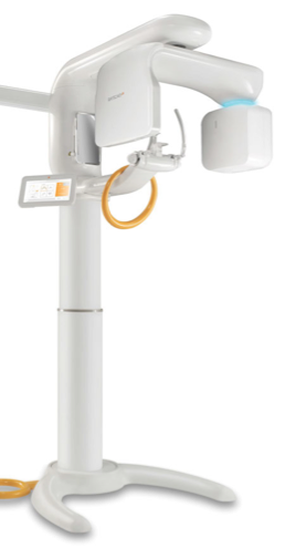 The Samsung RAYSCAN Alpha from LED Dental (www.leddental.com), for example, has an optimal 9 cm x 9 cm CBCT field of view that captures both jaws, including third molars for orthodontic treatment planning, and offers digital 2D panoramic and cephalometric capabilities. Developed by Ray Co., Ltd., a subsidiary of Samsung Electronics, the RAYSCAN Alpha is fully upgradable from 2D to 3D in-office, allowing practitioners to transition from film to digital, and ultimately, CBCT at their own pace. The system’s CMOS and Direct Deposition CsI Detector ensures practitioners can quickly capture high-quality, 16-bit DICOM images at a low radiation dose. To minimize errors from positioning and movement (a common issue with younger patients), the system’s focal trough is stabilized and controlled through Adaptive Moving Focus technology. The Samsung RAYSCAN Alpha – Expert’s proprietary noise reduction technology enhances image quality by removing noise that would otherwise blur images.
Due to its sleek design, the Samsung RAYSCAN Alpha – Expert can fit into nearly any practice’s floor plan. The system’s height adjustment component means patients of all sizes, including adolescents and patients in wheelchairs, can fit comfortably in the unit. The RAYSCAN Alpha – Expert is also the world’s first imaging system to utilize a wireless remote control for patient positioning. Additionally, green, blue, yellow, and red LED lights indicate the status of the unit to practitioners at a glance: ready, standby, exposure, and emergency.
In addition to the Samsung RAYSCAN Alpha – Expert’s advanced imaging capabilities and unique design, the system also features an intuitive user interface to streamline the capture process and simplify the manipulation of images. The unit also has a dedicated touch screen display for exposure parameter selection and image preview. The system utilizes LED Imaging Software for 2D images as well as Xelis 3D Imaging Software. With a 5-year warranty and the ability to integrate seamlessly with any practice management software, the Samsung RAYSCAN Alpha – Expert is a solid investment for any orthodontist.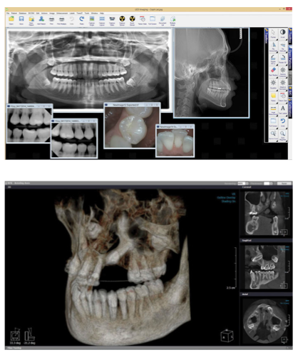
While 3D digital imaging systems are a larger investment than 2D imaging products, the time savings and return on investment for those cases requiring advanced 3D imaging will greatly benefit orthodontic practices in the long run. Ultimately, the best purchase clinicians can make for their practices is a multi-function unit with a long-term warranty from a trusted company that will be there before, during, and after the point of sale.
Since our launch in April, LED Dental has made a splash in the orthodontic market with 2D and 3D digital imaging products, highlighting our dedication to becoming one of the leading imaging providers in the industry. It’s our goal to provide practitioners with best-in-class imaging solutions ranging from extraoral imaging systems to intraoral cameras and beyond — so the next time you’re at a tradeshow, I invite you to come by our booth and explore the diverse product portfolio we have to offer!
This information was provided by LED Dental.
Stay Relevant With Orthodontic Practice US
Join our email list for CE courses and webinars, articles and mores
